Dr. James Stiehl, founder and inventor of the Perilav system, is still a treating clinician. He regularly sees patients in skilled nursing facilities and when appropriate uses the Perilav system as part of his treatment. He has done 1000’s of treatments and regularly documents the incredible results that he sees when Perilav is used in conjunction with effective topical wound management.
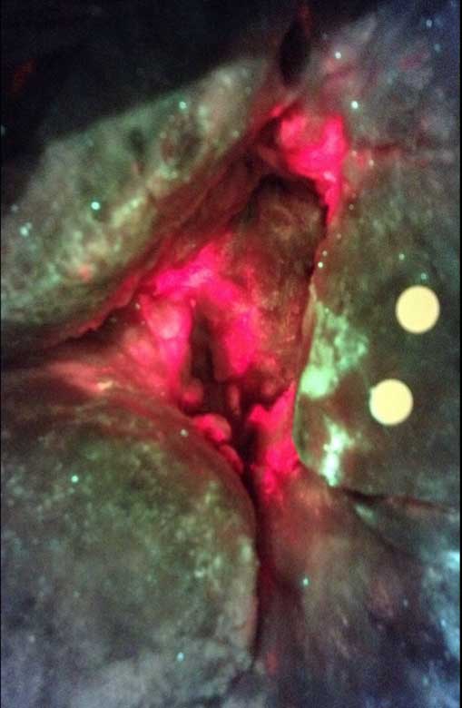
Before
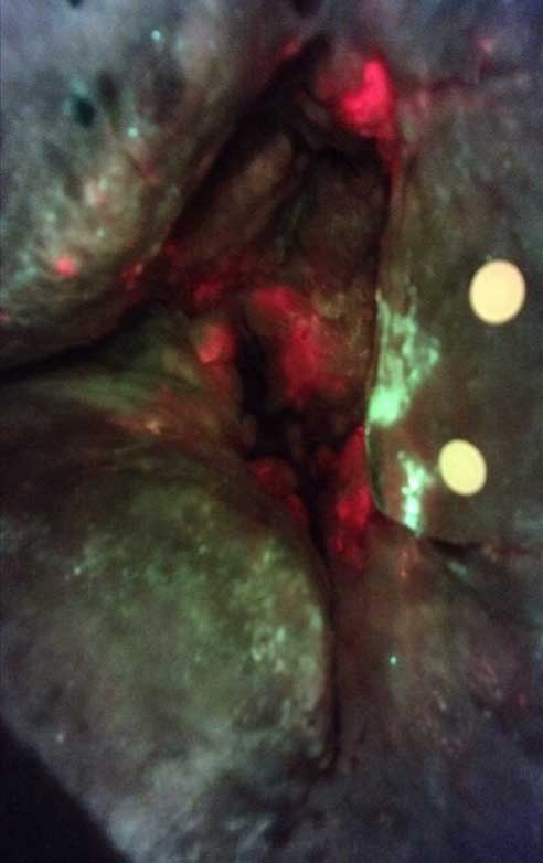
After
CASE STUDY #1
Wound assessed using MolecuLight autoflourescent imaging device. The red in the wound bed and the cyan on the periwound skin indicates bacterial levels >10^4 log. The wound was treated with Perilav using saline resulting in a significant reduction in the red areas within the wound.
Patient Age:
N/A
Wound type:
Sacral pressure injury
Previous treatment:
N/A
Frequency of Perilav treatment:
1
Time lapsed between photos:
20 min
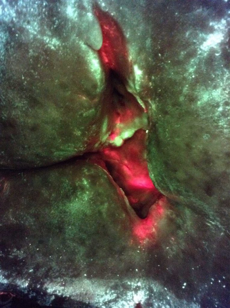
Before
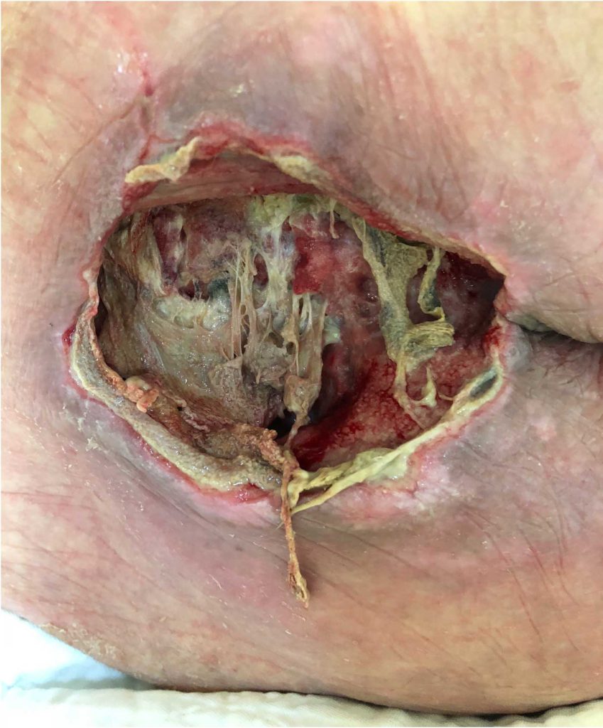
After
CASE STUDY #2
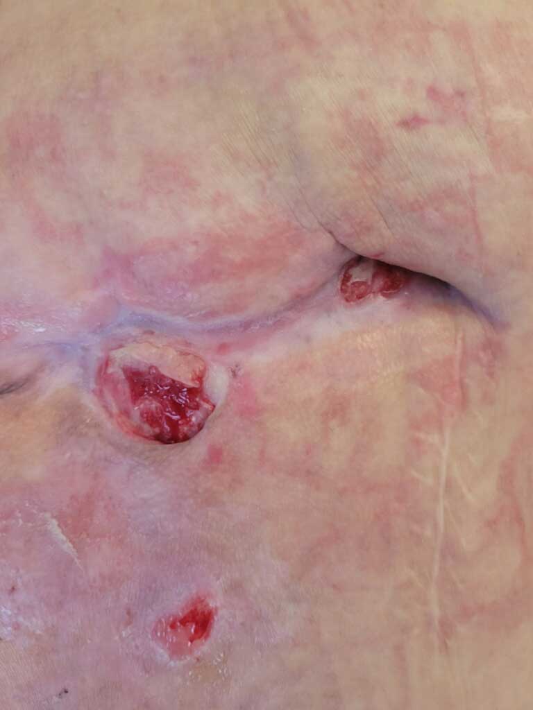
Sacral pressure injury assessed with MolecuLight prior to treatment. Red in the wound and cyan on the periwound indicate bacteria levels >10^4 log. Treated with Perilav using saline. Significant reduction of red areas in the wound.
Patient Age:
N/A
Wound type:
Sacral pressure injury
Previous treatment:
N/A
Frequency of Perilav treatment:
1
Time lapsed between photos:
15 min
Before
After
CASE STUDY #3
Patient returned to SNF after 10 day stay at the hospital with treatment of Santyl and NPWT.
Patient Age:
64
Wound type:
Sacral pressure injury
Previous treatment:
Santyl and NPWT
Frequency of Perilav treatment:
5x/week
Time lapsed between photos:
8 weeks
Before
After
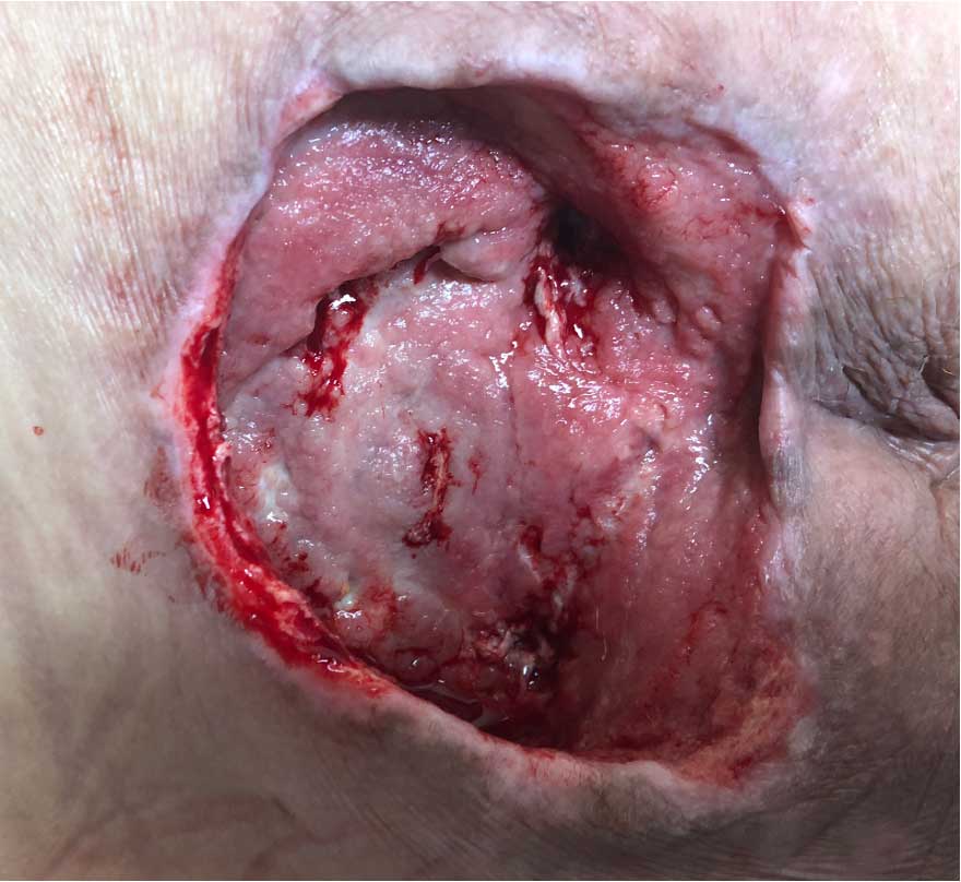
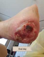
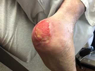
CASE STUDY #4
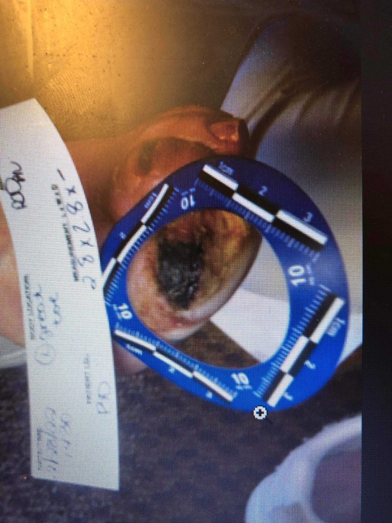
This patient had diabetic vascular disease, contralateral BKA, advanced chronic pulmonary disease, and insulin dependent diabetes. This was treated for 1 year with NPWT and this is the result.
Patient Age:
64
Wound type:
Heel Pressure Injury
Previous treatment:
Santyl and NPWT
Frequency of Perilav treatment:
3x/week
Time lapsed between photos:
6 weeks
Before
After
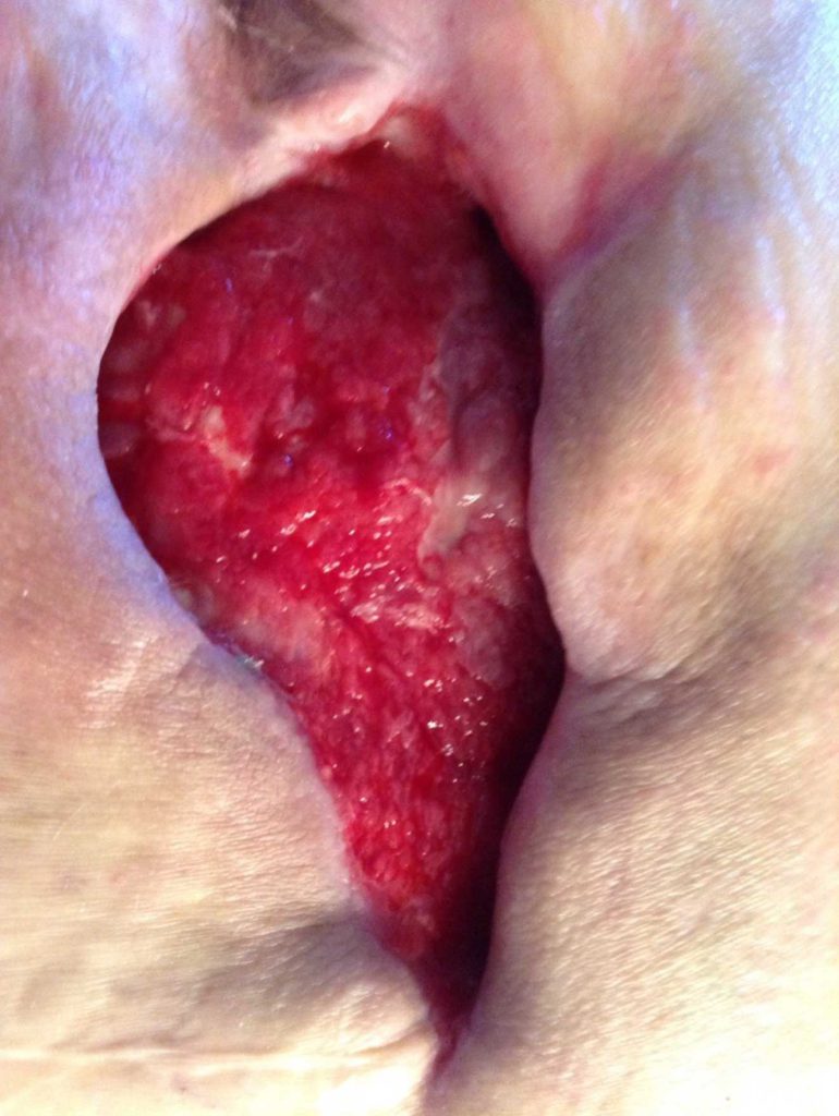
CASE STUDY #5
Sacral pressure injury. 243cm(2) including the undermined area. After only three treatments with Perilav.
Patient Age:
61
Wound type:
Osteomyelitis
Previous treatment:
NPWT x 8 weeks
Frequency of Perilav treatment:
3x/week
Time lapsed between photos:
7 weeks
Before
After
CASE STUDY #6
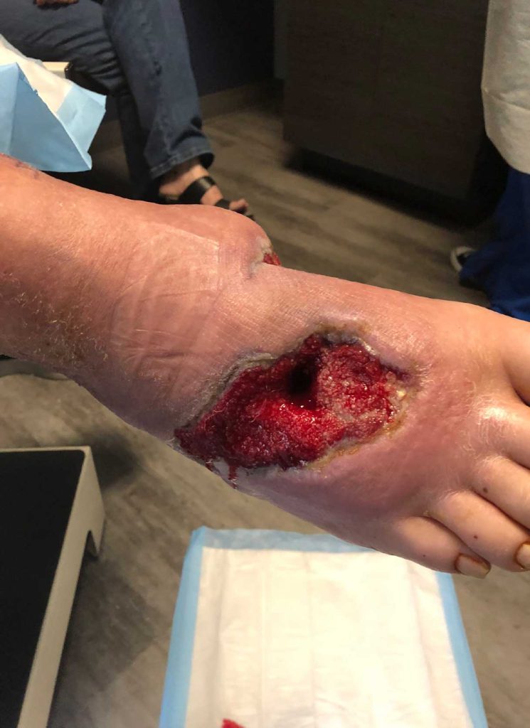
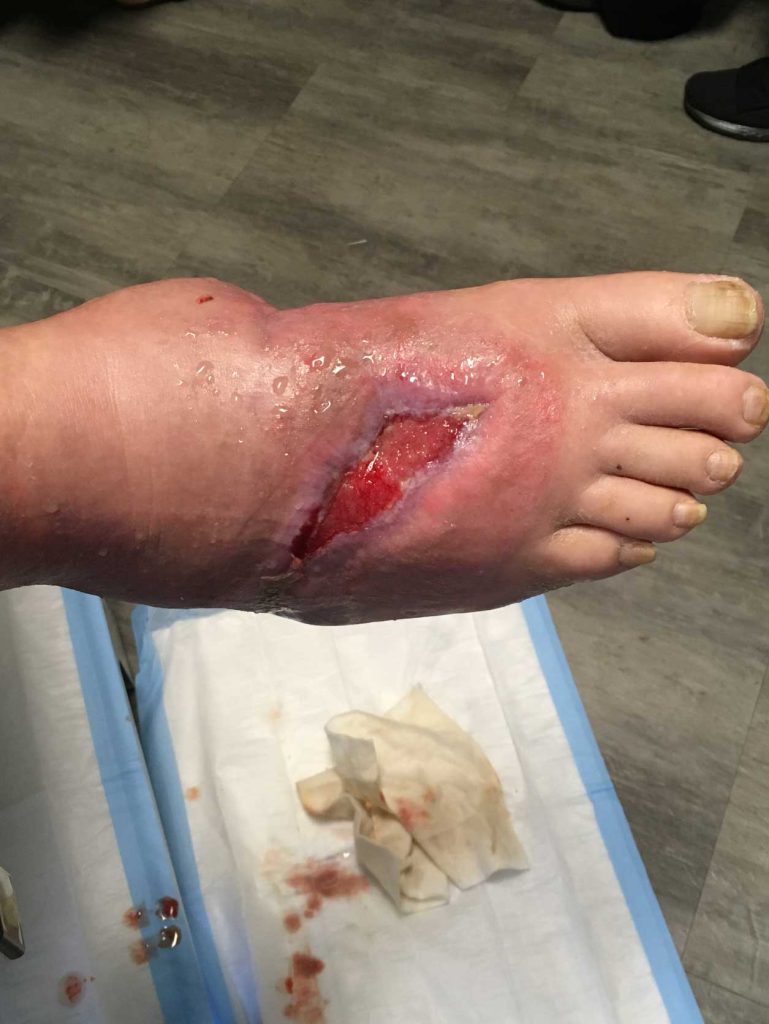
60 year old with mid foot infection (osteomyelitis) with diabetic Charcot foot. He refused BKA. Had a hind foot ulcer as well.
Patient Age:
60
Wound type:
Diabetic Foot Ulcer
Previous treatment:
8 weeks of NPWT
Frequency of Perilav treatment:
3x/wk x 6 weeks
Time lapsed between photos:
6 weeks
Before
After
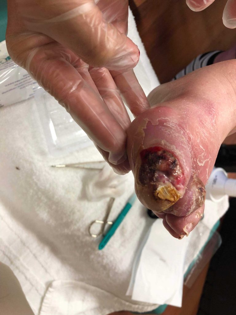
CASE STUDY #7
Patient with diabetic foot ulcer. One treatment with Perilav.
Patient Age:
82
Wound type:
Diabetic Foot Ulcer
Previous treatment:
N/A
Frequency of Perilav treatment:
1 treatment
Time lapsed between photos:
5 min